

🦷 Real Bone Graft Case: How We Preserve Bone After a Tooth Extraction (Part 1)
By Teuscher Legacy Dental – St. Charles & Campton Hills, IL
When most people think about tooth extractions, they imagine a simple “pull and heal.”
But modern dentistry allows us to preserve the bone where the tooth used to be — usually for setting the stage for a future dental implant that looks and functions like a natural tooth.
This post walks you through a real case from our practice showing what happens before, during, and right after bone grafting. There are different types of bone graft procedures- this one is technically a “socket preservation graft” because it “preserves” the “socket” where the tooth used to be, by filling it with bone. So for this post that’s the type of graft we mean if we say “bone graft”.
In part 1, we will show and discuss the actual extraction and bone graft procedure. In Part 2, we’ll show how it healed and how we planned the dental implant using 3D CBCT imaging.
But First: should you even get a bone graft? Why do you need one? When is it OK to not have one?
I’ll give more detail in a future post. But below is a little more information. The short story is, socket preservation isn’t mandatory for every extraction — but it’s usually the smartest long-term choice if you plan to replace the tooth with an implant.
Here’s how the benefits and risks of socket preservation grafts compare:
| Decision | Benefits | Possible Risks / Costs |
|---|---|---|
| Perform Bone Graft (Socket Preservation) |
|
|
| Skip Bone Graft (No Preservation) |
|
|
Sources: Araujo & Lindhe 2005; Misch 2015; Pikos MA 2019; Kois Center Clinical Protocols.
👀 Step 1: Pre-Op Evaluation
Before removing the tooth, we assessed bone health and root position with detailed imaging. In this case, the tooth had a prior root canal and a cavity on the back side, which caused the crown to loosen and eventually fall off. This tooth could not be saved by a new crown, so it needed to be extracted to prevent risk of infection and further damage of surrounding tissue. After reviewing alternative options, this patient is planning on a future dental implant to restore the site.
Images:

X Ray of tooth #18 Prior to Extraction

Photo of tooth #18 prior to extraction
✅ Purpose: understand root formation, nerve anatomy, and ensure the socket walls are intact for grafting.
🦷 Step 2: Tooth Extraction
After gentle removal, the socket (the bony hole where the roots sat) is clearly visible.
Left alone, this socket starts to shrink immediately — studies show 30–60 % of ridge width can disappear within the first year after extraction (Araujo & Lindhe, Clin Oral Implants Res, 2005). That bone loss often makes future implants more difficult and can even affect facial contours.
Image:
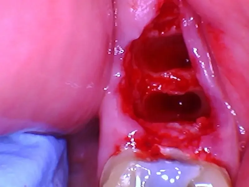
The socket after gentle extraction.
*note for the adjacent tooth - we also did crown #19 that day, and this was before we cemented the temporary crown.
✅ Goal: Gentle extraction maintains socket integrity, keeping bone walls present, and prevents early bone collapse.
🧪 Step 3: Bone Graft and Collagen Plug
To prevent bone collapse, we placed a biocompatible bone graft into the socket.
The material acts as a scaffold that your body gradually replaces with living bone. Usually these bone grafts are from human cadavers, (allograft) but can also be from your own body (autograft), cows or horses (xenograft), or completely synthetic (alloplast).
In our case, we used a cadaver bone particle mix to fill the socket. A collagen plug and PTFE non-resorbable sutures were added to stabilize the area and protect the graft during healing.
Images:

A clear picture of the collagen plug over the top of the bone graft. You can see two of the white sutures as well.

After the bone graft. Notice where the roots used to be, the socket appears filled with bone particulate.
✅ Goal: preserve ridge volume, gum contour, and future implant site quality.
From here, the body takes over — blood flows into the bone graft and forms a clot- eventually forming new bone inside the socket over the next several weeks.
⏱ What’s Next
In Part 2, we’ll show:
The one-week post-op appearance
The 8-week CBCT scan showing regenerated bone
How we plan precise implant placement using guided surgery
💬 Why We Share Cases Like This
We believe you deserve transparency.
Seeing the actual process (not just a before-and-after) helps you understand why bone preservation matters and what is actually happening in the bone graft procedure.
Once bone is lost, it’s more expensive and less predictable to rebuild. Preserving it early gives you more control, fewer surgeries, and a more natural final result.
📍 About Teuscher Legacy Dental
St. Charles & Campton Hills, IL
Family-run, fee-for-service practice specializing in advanced surgical and cosmetic dentistry.
🏆 Voted Best Cosmetic Dentist in St. Charles 2025 by Business Rate.
Learn more about:
🔜 Coming Soon: Part 2
“How Bone Grafts Heal: 8-Week CBCT Results and Implant Planning”
We’ll show real post-op images, bone density changes, and how careful socket preservation makes precise implant placement possible.